Neutron Imaging
Similar to x-ray radiography, neutron radiography is a very efficient tool to enhance investigations in the field of non-destructive testing (NDT), as well as in many fundamental research applications. Neutron radiography is, however, suitable for a number of tasks impossible for conventional x-ray radiography. The advantage of neutrons compared to x-rays is the ability to image light elements (i.e. with low atomic numbers) such as hydrogen, water, carbon, etc. In addition, neutrons penetrate heavy elements (i.e. with high atomic numbers) such as lead, titanium, etc. allowing the study of materials in complex sample environments; for example, water accumulation in hydrogen fuel cells. Because neutrons interact with the nucleus rather than with the electron shell, they can also distinguish between different isotopes of the same element. This makes neutron radiography an important tool in various research applications and in the field of NDT. The high neutron intensity beams permit short exposure times, high spatial resolution, and high sample throughput.
MNRC offers the largest neutron radiography bays in the world, allowing samples to be imaged ranging in size from less than 1 cm in length and 1 gram in weight up to several meters in length and over 1,000 kilograms.
For Neutron Radiography inquiries, please refer to the Contact Us page.
Methods of Neutron Imaging
The detection of neutrons relies on a conversion into visible light; to achieve this, conversion screens containing either Gd or 6Li and a fluorescence material are commonly used. After the conversion, the emitted light can be detected by different media such as:
- Film, that is then developed in a dark room and results in a permanent image.
- Imaging Plates, that can be re-used after being processed by an image reader. (The technology is very similar to x-ray imaging plates used at medical offices).
- Digital cameras (CCD, CMOS), allow to capture the image digitally.
The resolution of all systems is approximately 100 microns.
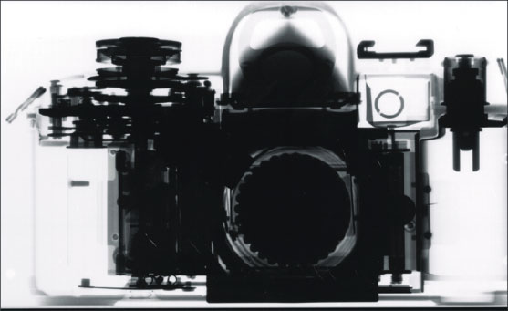
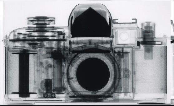
Differences Between Neutron and X-ray Radiography
Neutron radiography is based on the principal that neutrons interact with the nucleus of the atom, rather than the electrons. Therefore, neutrons are absorbed in matter very differently from x-rays and gamma rays. This means that, contrary to x-rays, neutrons are attenuated by some light materials, such as hydrogen, boron, and lithium, but penetrate many heavy materials such as titanium and lead. Penetration depth can range up to several inches, allowing for some unique applications of neutron radiography.
The images below demonstrate how neutron radiography can yield different, yet complementary information to x-ray radiography.